
Zebrafish Light Sheet Fluorescence Microscopy
Vascular diseases are the leading cause of death world-wide. Our understanding of the mechanisms that govern vascular development and diseases processes rely almost completely on in vitro and preclinical in vivo models.
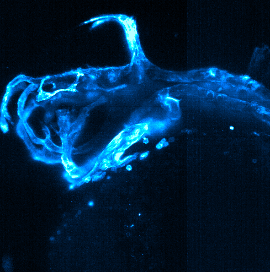
Zebrafish are an established model to study vascular development and disease in vivo, over time, and in three dimensions (3D). Embryonic transparency, coupled with availability of various transgenic lines, and rapid development makes zebrafish an unrivalled model for 3D in vivo imaging.
Light sheet fluorescence microscopy (LSFM) is a microsopy technique that allows optical sectioning by an uncoupling of the excitation and emission beam, reducing photobleaching and increasing imaging speed.
Together, zebrafish transgenic lines and LSFM allows for the study of vascular development and disease with unprecedented detail over long periods of time (hours-days).
During her PhD, Elisabeth used the commercial Zeiss Z.1 LSFM to acquire data for her quantification as well as kugeln project. Her images were featured on various covers and webpages. Based on her expertise, she has given several presentations on LSFM, regularly trains and advises biologist on experimental design.
Publications
Spotlight - Barresi M. #, Coen E. #, Kugler E. #, Shuda J. #, and Sung D.#, Engaging new audiences with imaging and microscopy.
Development 15 September 2021; 148 (18): dev199942. doi: https://doi.org/10.1242/dev.199942
Journal Article - Kugler E.*, Snodgrass R., Bowley G., Plant K., Serbanovic-Canic J., Hamilton N., Evans P., Chico T., and Armitage P.,
The effect of absent blood flow on the zebrafish cerebral and trunk vasculature. Vascular Biology, Online Publication Date: 25 Aug 2021 https://doi.org/10.1530/VB-21-0009
Review - Bowley G. #, Kugler E. #, et al., Zebrafish as a tractable model of human cardiovascular disease, British Journal of Pharmacology, 2021, https://doi.org/10.1111/bph.15473
Review - Chico T.* and Kugler E.*, Cerebrovascular development: mechanisms and experimental approaches, Cell. Mol. Life Sci., Mar. 2021, doi: 10.1007/s00018-021-03790-1. (Link)


